Guard Cells (Cambridge International A-Level Biology)
Spec point —
14.2.3· describe the structure and function of guard cells and explain the mechanism by which they open and close stomata
Guard cells
Structure of guard cells
-
Each stoma is surrounded by two guard cells
-
Guard cells have the following features:
- Thick cell walls facing the air outside the leaf and the stoma
- Thin cell walls facing adjacent epidermal cells
- Cellulosemicrofibrils arranged in bands around the cell
- Cell walls have no plasmodesmata
- Cell surface membrane is often folded and contains many channel and carrier proteins
- Cytoplasm has a high density of chloroplasts and mitochondria
- Chloroplasts have thylakoids but with few grana (unlike those in mesophyll cell chloroplasts)
- Mitochondria have many cristae
- Several small vacuoles rather than one large vacuole
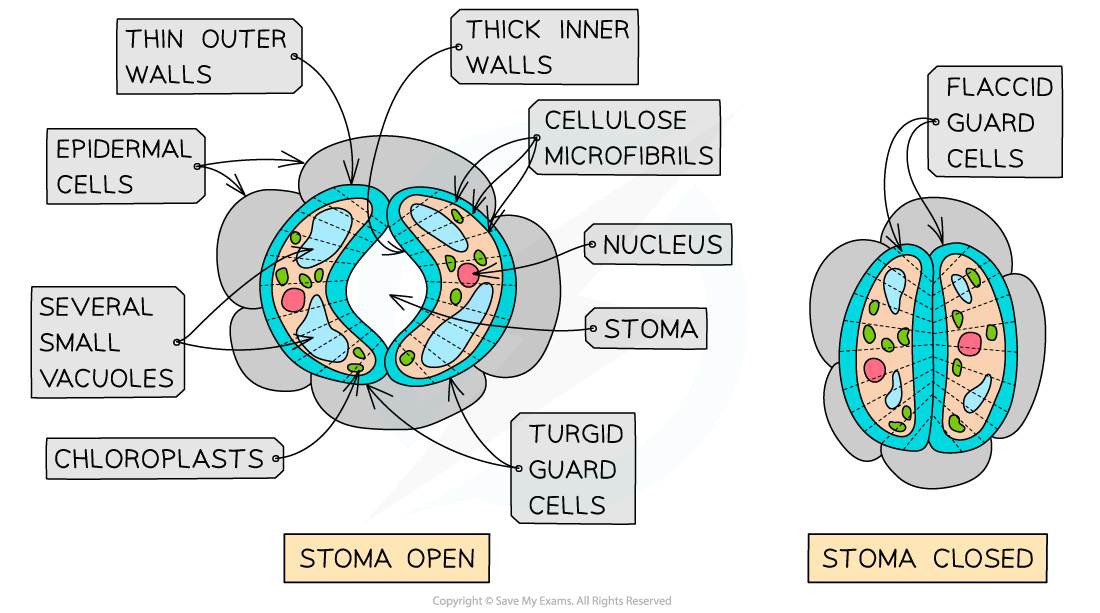
The structure of guard cells when the stoma is open and closed
Mechanism to open stomata
-
Guard cells open when they gain water and become turgid
-
Guard cells gain water by osmosis
-
A decrease in water potential in the guard cells is required for water to enter the cells by osmosis
-
In response to light, ATP-powered proton pumps in the guard cell surface membranes actively transport hydrogen (H+) ions out of the guard cell
-
This leaves the inside of the guard cells negatively charged compared to the outside
-
This causes channel proteins in the guard cell surface membranes to open, allowing potassium (K+) ions to move down the electrical gradient and enter the guard cells
-
The potassium (K+) ions also diffuse into the guard cells down a concentration gradient
- The combination of the electrical gradient and concentration gradient is known as an electrochemical gradient
-
The influx of potassium (K+) ions increases the solute concentration inside the guard cells, lowering the water potential inside the cells
-
Water now enters the guard cells by osmosis through aquaporins in the guard cell surface membranes
- Most of the water enters the vacuoles, causing them to increase in size
-
This increases the turgor pressure of the guard cells, causing the stoma to open
- The bands of cellulose microfibrils only allow the guard cells to increase in length (not diameter)
- The thin outer walls of the guard cells bend more easily than thick inner walls
- This causes the guard cells to become curved, opening up the stoma
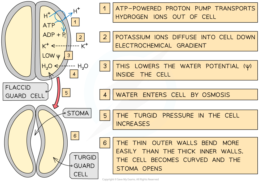
An outline of the mechanism to open stomata
Mechanism to close stomata
- When certain environmental stimuli are detected (that lead to the closing of the stomata), the proton pumps in the guard cell surface membranes stop actively transporting hydrogen (H+) ions out of the guard cell
- The potassium (K+) ions leave the guard cells
- The water potential gradient is now reversed and water leaves the guard cells by osmosis
- This causes the guard cells to become flaccid, closing the stoma
Was this revision note helpful?
Build on this topic