Recognising Tissues (Cambridge International A-Level Biology)
Recognising Tissues in the Gas Exchange System
Spec point —
9.1.3· recognise cartilage, ciliated epithelium, goblet cells, squamous epithelium of alveoli, smooth muscle and capillaries in microscope slides, photomicrographs and electron micrographs
Cartilage
- The cartilage in the trachea has a ‘glassy’ appearance due to its translucent protoplasm
- It contains no nerves or blood vessels
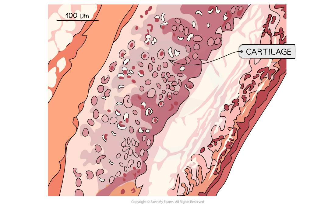
Photomicrograph of cartilage tissue in the gas exchange system
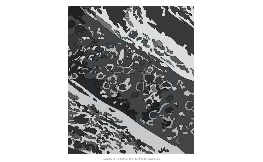
Electron micrograph of cartilage tissue in the gas exchange system
Ciliated epithelium
-
Ciliated epithelial cells are distinctive due to their:
- Narrow cell bodies
- Hair-like cilia located along the apical layer
-
The cilia are tiny hair-like projections that beat to sweep mucus (with trapped dust and pathogens) up and away from the lungs
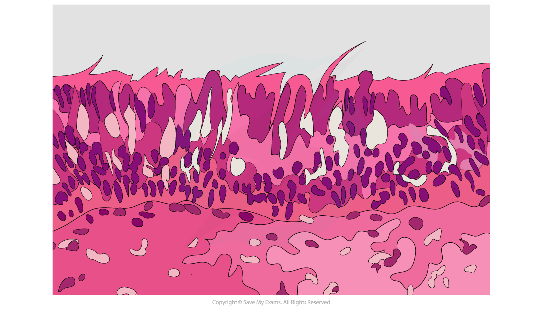
Photomicrograph of ciliated epithelium tissue in the gas exchange system
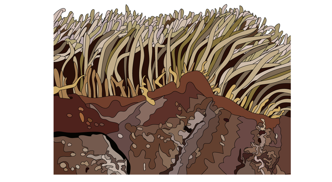
Electron micrograph of ciliated epithelium tissue in the gas exchange system
Goblet cells
- Goblet cells can be found scattered among the ciliated epithelium of the trachea and bronchi
- They are distinctive in the epithelium due to their lack of cilia
- The nucleus is found towards the basal (bottom) layer of the cell, with a large Golgi apparatus and mucus granules found towards the apical layer
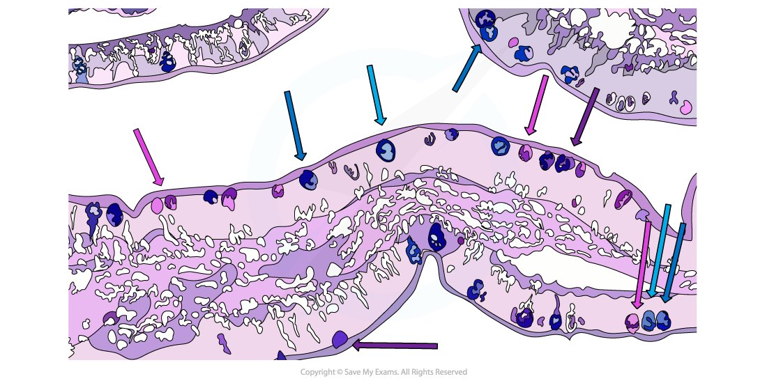
Photomicrograph of goblet cells in the gas exchange system
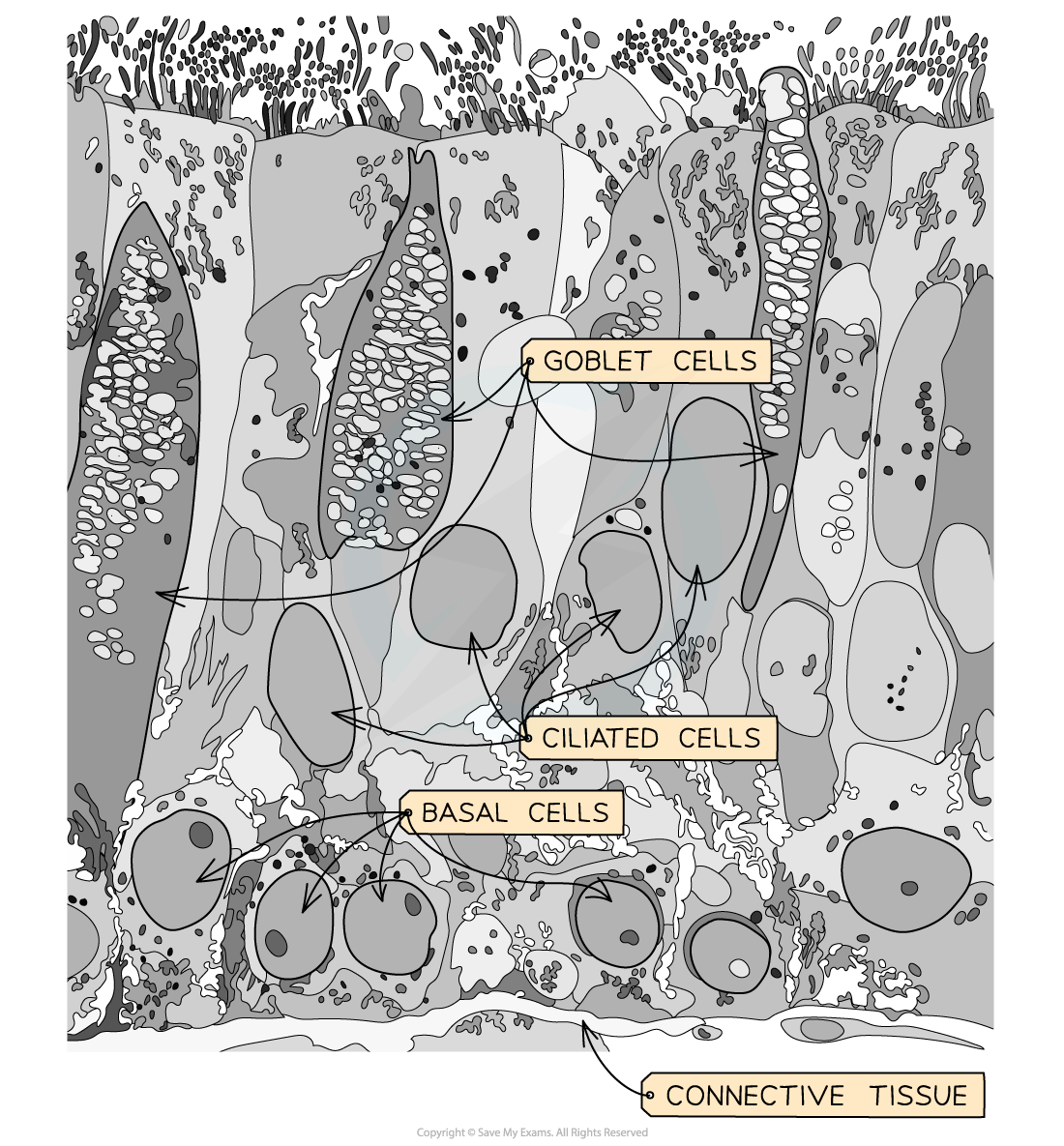
Electron micrograph of goblet cells in the gas exchange system
Squamous epithelium of alveoli
- Squamous epithelium is made of thin, flat squamous cells
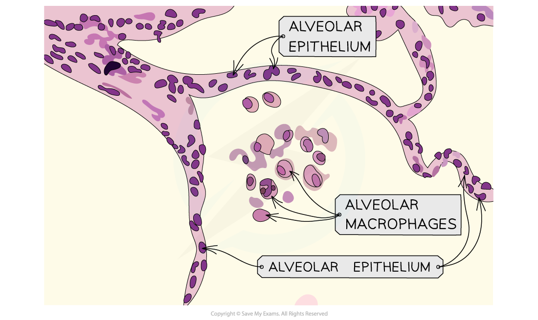
Photomicrograph of squamous epithelium in the gas exchange system
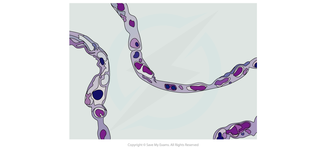
Electron micrograph of squamous epithelium in the gas exchange system
Smooth muscle
- The cells of smooth muscle are tightly packed and are found beneath the ciliated epithelium
- Unlike skeletal muscles, they are not striated and so don’t show any cross stripes under a microscope
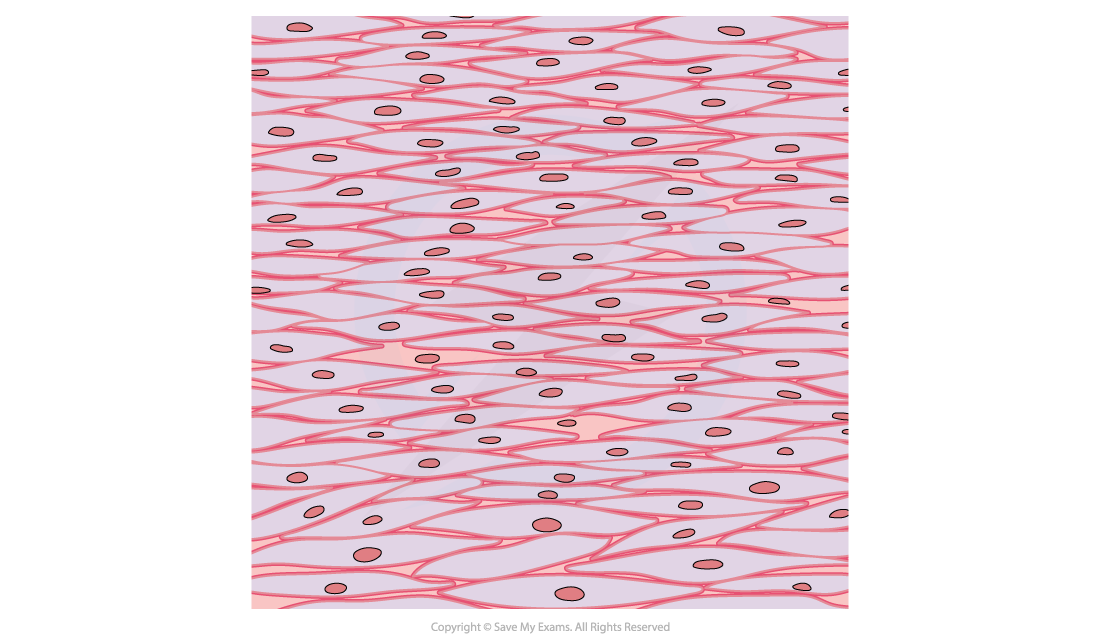
Photomicrograph of smooth muscle in the gas exchange system
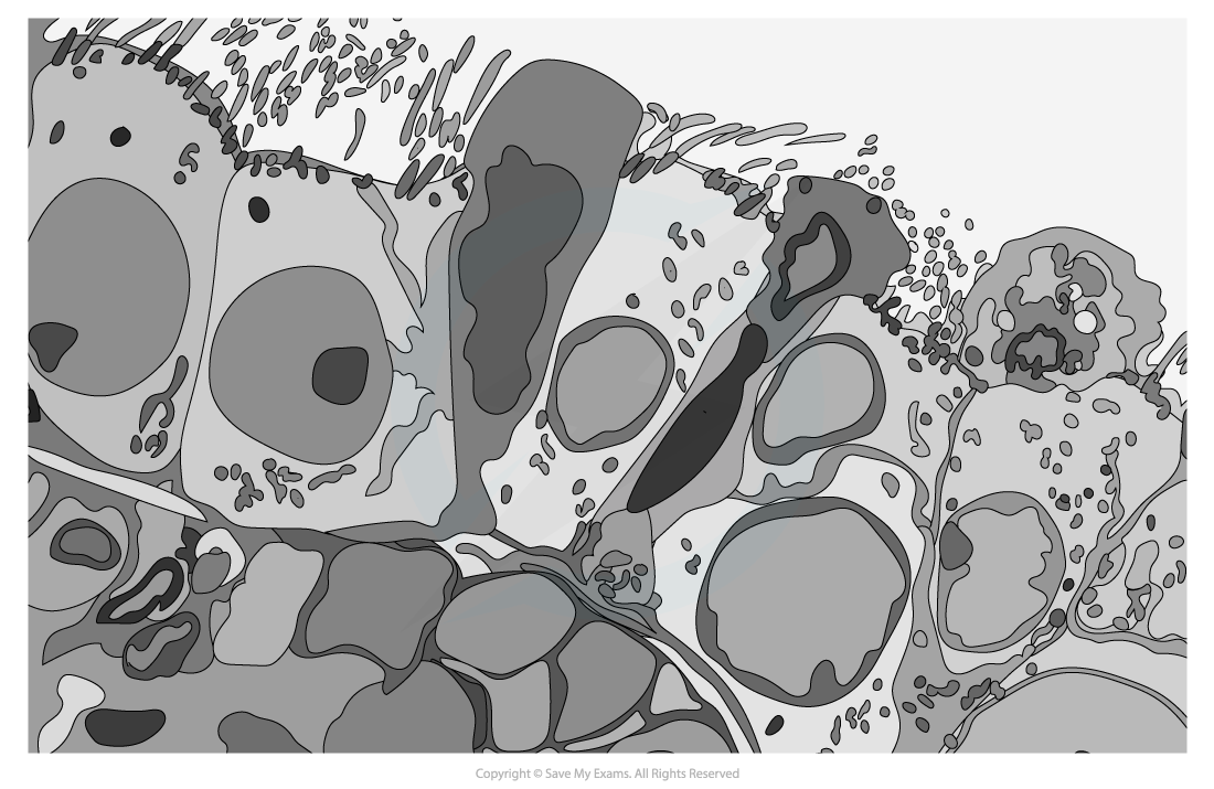
Electron micrograph of smooth muscle in the gas exchange system
Capillaries
- Capillaries are distinctive from other blood vessels due to their tiny diameter (about 7 μm, roughly one red blood cell wide)
- Their walls consist of a single layer of epithelial cells
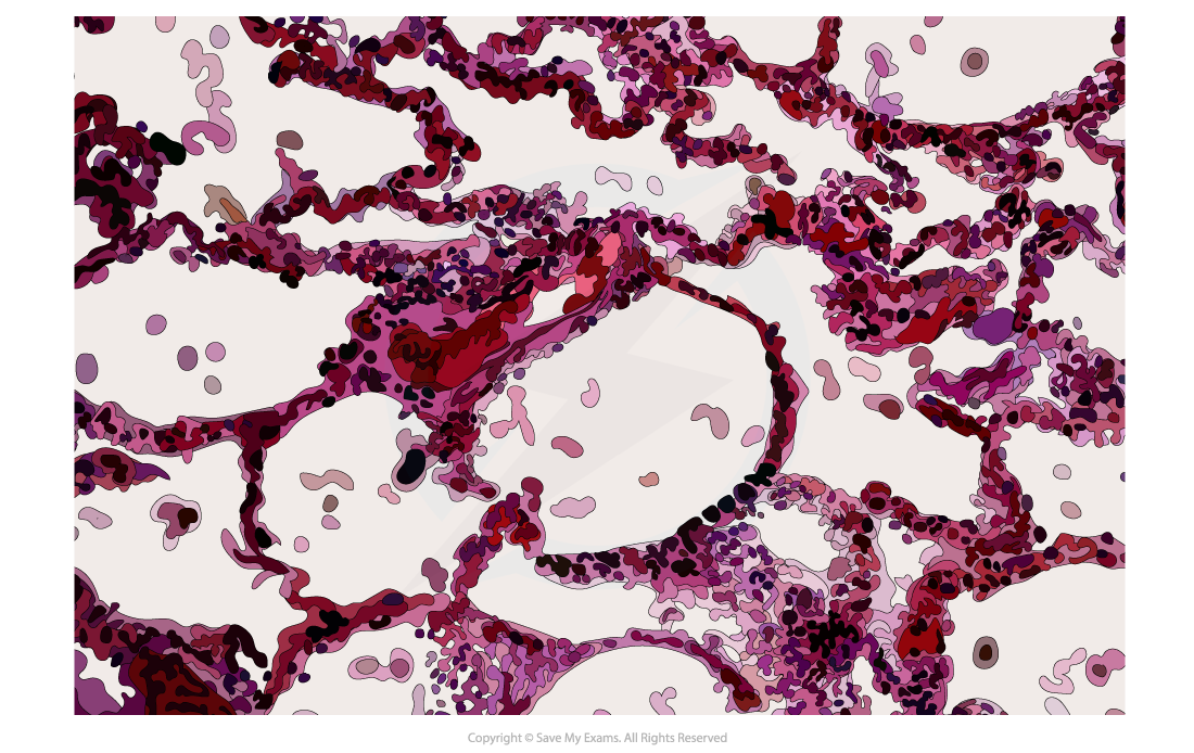
Photomicrograph of capillaries in the gas exchange system
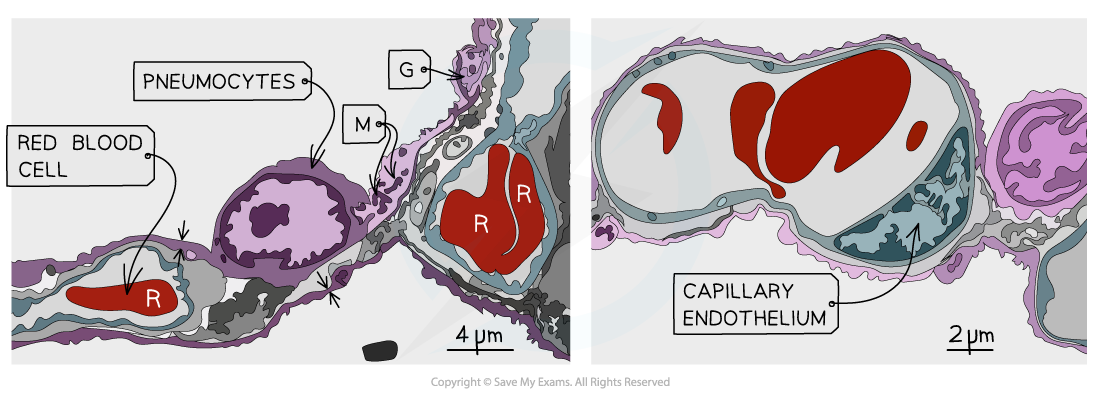
Electron micrograph of capillary tissue in the gas exchange system
Was this revision note helpful?
Build on this topic